|
|
Triplane Fracture
General Considerations
- The fracture involves the frontal, lateral, and transverse planes
- It extends sagittally through the epiphysis, transversely through the epiphyseal plate and coronally through the distal tibial metaphysis
- Occurs in older children/young adolescents in 18 months just prior to epiphyseal plate closure (about 12-15 years)
- This is related to the manner in which the distal tibial epiphyseal plate closes
- Children are susceptible to triplane fractures after closure of the medial epiphyseal plate and before closure of the lateral epiphyseal plate
- Mechanism of injury is forced external rotation
- They may be classified by the number of fragments into two-part, three-part and four-part triplane fractures
- Two-part fracture is most common
- In classic lateral two-part fracture, a posterolateral fragment, consisting of the posterolateral epiphysis and the posterior metaphysis, separates from the anteromedial epiphysis, which remains attached to the tibial shaft
- In the classic medial three-part fracture, there is a free anterolateral epiphyseal fragment, a fragment involving the rest of the epiphysis and the posterior metaphysis, and the main fragment corresponding to the rest of the tibial metaphysis and shaft
Clinical Findings
- Localized pain
- Swelling
- Inability to fully bear weight
- Ankle deformity
Imaging Findings
- Conventional radiography is the study of first choice
- CT is helpful in optimally demonstrating all of the fracture fragments and their relationships
- Two-part fracture appears on lateral view as if it were a Salter-Harris IV fracture
- Three-part fracture appears as if it were a Salter-Harris III fracture on AP view and as if it were a Salter-Harris II fracture on lateral view
Differential Diagnosis
- Tillaux fracture - Salter-Harris III fracture involving the distal tibial epiphysis
Treatment
- Most non-displaced, two-part triplane fractures can be treated by closed reduction
- Displaced fractures and most three-part triplane fractures require operative reduction and internal fixation
Complications
- Spiral fracture of fibula occurs in 50% of patients
- Epiphyseal arrest and angular deformity are uncommon

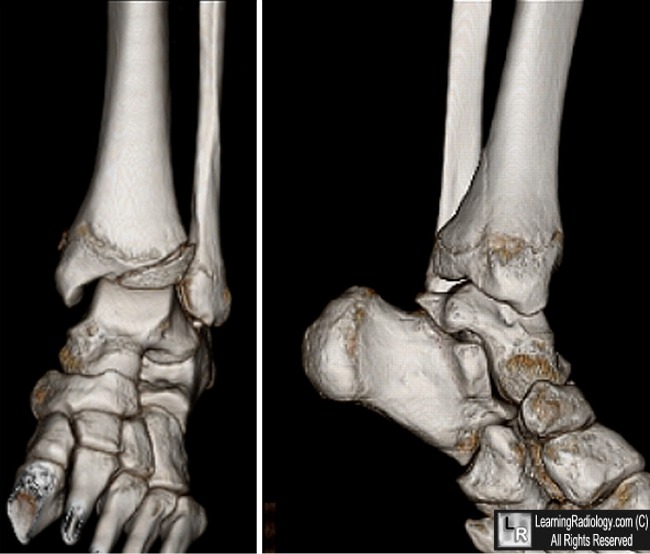
Triplane Fracture of Tibia.On the AP (coronal) view on left, the fracture line extends through the epiphysis (blue arrow), laterally across the epiphyseal plate (yellow arrow). On the lateral (sagittal) view on the right, the fracture line extends through the epiphysis and into and out of the metaphysis (red arrows).
For this same photo, click here
For more information, click on the link if you see this icon 
Analysis of 51 Tibial Triplane Fractures Using CT with Multiplanar Reconstruction.SD Brown, JR Kasser, D Zurakowski and D Jaramillo. AJR 2004;183:1489–1495
Wheeless Online
Triplane Fracture. J Abt. eMedicine
|
|
|
